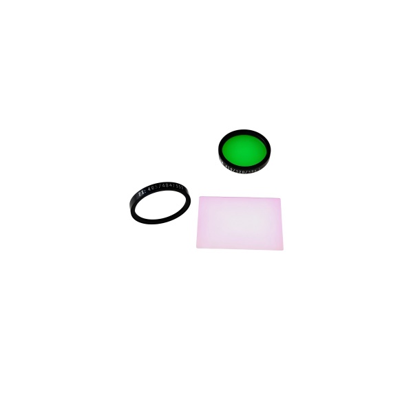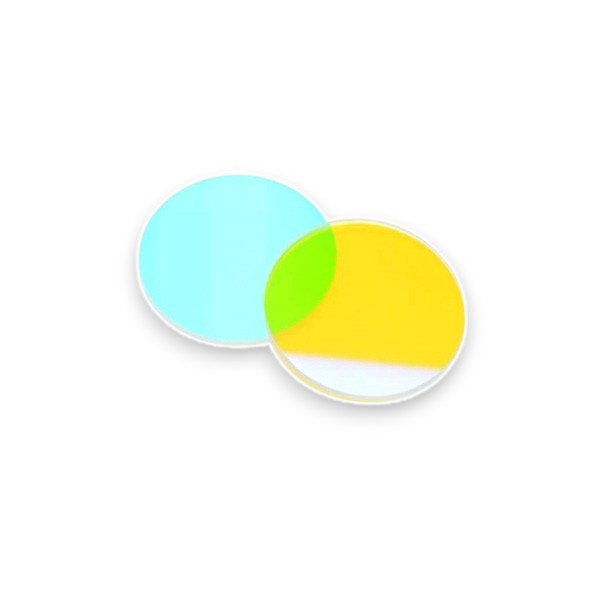
415.5nm Dichroic Optical Filter
415nm dichroic optical filter is designed to reflect light at 415.5nm and transmit longer wavelength. Coligh manufactures high precision dichroic mirror for fluorescence microscopy and imaging system.
- Precise Reflective Wavelength at 415.5nm
- 90%Excellent Transmission Performance
- Hard Coating
- Steep Transition
- 45° angle of incidence
Products Categories
Get A Free Quote
Narrow Edge 415.5nm Longpass Dichroic Filter Description
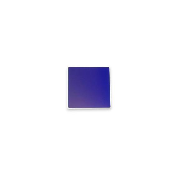
415.5nm dichroic optical filter Coligh manufactures is a spectroscopic device based on the principle of optical interference. The 415nm dichroic filter can selectively transmit the 415.5nm wavelength band according to the wavelength, and reflect and block the ultraviolet or near-ultraviolet light.
- The edge wavelength is 415.5+/-1nm, specially designed for the ultraviolet-visible light boundary, suitable for mercury lamp spectrum or ultraviolet laser
- The reflectivity of the band less than 415.5nm is greater than or equal to 98%
- The transmittance of the band greater than 420nm is greater than or equal to 90%, minimizing the loss of light energy.
- We use ion-assisted deposition hard coating. We deposit high-refractive index and low-refractive index dielectric films on fused quartz. The dichroic filter is resistant to high temperature and UV radiation aging, suitable for industrial and outdoor scenes.
415.5nm Dichroic Optical Filter Technical Datasheet
| Parameter | Specification |
| Product Type | Dichroic Optical Filter / Longpass Beamsplitter |
| Edge Wavelength | 415.5 nm ± 2 nm |
| Angle of Incidence (AOI) | 45° |
| Reflectance | ≥98% |
| Transmission Range | > 90% average |
| Working Wavelength Rang | 350-650nm |
| Coating Type | Hard-coated dielectric multilayer |
| Substrate Material | Fused Silica |
| Surface Quality | 60/40 or better (per MIL-PRF-13830B) |
| Size Options | 15*11*1.0mm, custom sizes available |
| Clear Aperture | > 90% of dimension |
| Thickness | 1.0 mm ± 0.1 mm |
415.5nm Dichroic Optical Filter Applications
- Fluorescence microscope
Multicolor fluorescence imaging requires simultaneous excitation of UV DAPI and visible light dye FITC, but UV excitation light can easily contaminate the visible light detection channel. The 415nm dichroic filter is placed in the microscope light path at a 45° tilt angle to reflect UV light to the sample and transmit visible fluorescence signals to the camera. The UV light is precisely directed to the sample to excite the DAPI-stained cell nucleus. Blue fluorescence is allowed to pass while blocking UV reflected light from entering the detector. This eliminates the need to switch filters, improving imaging speed and multi-task detection capabilities. - Flow cytometer
The 355nm UV + 488nm blue multi-laser system requires spectrometry to avoid crosstalk, but traditional spectroscopes are large and inefficient. The 415nm dichroic mirror can be integrated into the laser beam combining module to reflect the UV laser to the sample flow and transmit the blue laser to another optical path. : Ensure that each laser independently excites different fluorescent dyes - Laser processing system
The 355nm UV laser and the visible light positioning laser 635nm red light need to share the same optical path, but the UV light protection sensor needs to be isolated. The 415.5nm dichroic mirror can be installed between the galvanometer and the workpiece to reflect the UV processing laser to the processing area and transmit the visible positioning light to the visual calibration system. It blocks the UV laser from damaging the CCD camera and only transmits visible light for real-time monitoring.